Retinal Detachment
Definition
Detachment of retinal from choroid plexus, resulting in no perfusion of outer 1/3 of retina (RPE ++)
NOTE: Ophthalmic emergency!! Refer immediately
Types:
Epidemiology
Risk Factors
Detachment of retinal from choroid plexus, resulting in no perfusion of outer 1/3 of retina (RPE ++)
NOTE: Ophthalmic emergency!! Refer immediately
Types:
- Rheumatogenous (aka break) - full thickness tear. Cause: pre-existing hole, trauma, surgery, PVD (causes a break)
- Tractional. Cause: PDR --> retinal membrane contract
- Exudative/ haemorrhagic (less common). Cause: Tumour, inflammatory disease, congenital anomalies of blood-retina barrier
Epidemiology
- 1/10,000
- M=F
- 35% post eye op
Risk Factors
- Myopia (large eyeball, thin retina)
- Eye surgery
- Trauma
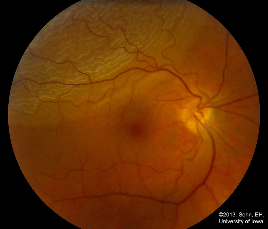
Retinal Detachment.
Clinical Features
Symptoms
Signs
Management
If macula still attached, immediate surgery
If macula detached, may be able to wait, depending on ophthalmologist's call
Source
www.ophtobook.com 2009
Symptoms
- Flashes (aka photopsia) and floaters (common)
- Dark curtain (less common)
Signs
- Fundoscope examination
- Shafer's sign = pigmented particle (RPE) in anterior chamber = pathognomonic for retinal tear
Management
If macula still attached, immediate surgery
If macula detached, may be able to wait, depending on ophthalmologist's call
- Vitrectomy
- Pneumatic retinopexy
- Scleral buckling (belt around anterior chamber)
- Laser pegging (prophylaxis to prevent detachment)
Source
www.ophtobook.com 2009
