
Pulmonary Embolus (PE)
Summary
Symptoms
Compression stockings --> O2 --> Morphine --> Thrombolysis if critical --> Clexane
Symptoms
- Sharp chest pain
- Dyspnoea
- Haemoptysis
- Pre/ syncope
- Leg pain
- Immobility
- Oestrogen therapy
- Surgery
- Cancer
- PMHx
- FHx
- Cyanotic
- Tachycardia
- HI JVP, loud P2, RV heave
- Reduced breath sounds
- (Calf tenderness)
- Bedside: ECG
- Bloods: D-dimer (rule out), ABG, coags, FBC+CRP, UEC, troponin, BNP
- Imaging: CTPA/MRPA (gold), V/Q scan, CXR, Doppler US of legs (DVT), 2D Echo (heart emboli)
Compression stockings --> O2 --> Morphine --> Thrombolysis if critical --> Clexane
- (If SBP >90) --> warfarin loading
- (If SBP <90) --> colloid infusion --> dobutamine --> IV noradrenaline --> thrombolysis/ emoblectomy
Definition
Emboli lodged in pulmonary artery/ arterioles
Aetiology
Most commonly dislodged thrombus from proximal legs
Risk Factors
Clinical Features
Symptoms
Investigations
Bedside: ECG (tachy, S1Q3T3, T wave inversion V1-4)
Bloods: D-dimer (non specific = rule out), BNP (myocardial stretch), troponin (RV infarct), ABG (hypervent/ poor gas exchange), FBC+CRP (baseline)
Imaging: CTPA (gold standard), MRPA (for renal insufficiency), lung V/Q scan (no contrast needed), CXR (often normal/ P.effusion/ Atelectasis), Doppler US of legs (DVT), 2D Echo (heart emboli)
Diagnosis
Emboli lodged in pulmonary artery/ arterioles
Aetiology
Most commonly dislodged thrombus from proximal legs
Risk Factors
- Immobility
- Oestrogen therapy
- Surgery
- Cancer
- PMHx
- FHx
Clinical Features
Symptoms
- Pleuritic chest pain
- Dyspnoea
- Haemoptysis (breakdown of lung tissue)
- Pre/ syncope
- Cyanotic
- Fever
- Tachycardia
- HI JVP, loud P2, RV heave
- Reduced breath sounds
- (Calf tenderness/ swelling)
Investigations
Bedside: ECG (tachy, S1Q3T3, T wave inversion V1-4)
Bloods: D-dimer (non specific = rule out), BNP (myocardial stretch), troponin (RV infarct), ABG (hypervent/ poor gas exchange), FBC+CRP (baseline)
Imaging: CTPA (gold standard), MRPA (for renal insufficiency), lung V/Q scan (no contrast needed), CXR (often normal/ P.effusion/ Atelectasis), Doppler US of legs (DVT), 2D Echo (heart emboli)
Diagnosis
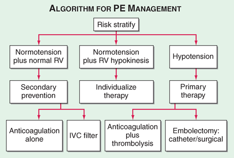
Management
Prevention!
Acute Mx:
Prevention!
- Compression stockings
- VTE prophylaxis (LWMH)
Acute Mx:
- O2
- Fluids
- Analgesia
- (Below)
Complications
RVF
Bleeding risks from anticoagulants (HIT)
Source
Harrison's 18th Ed 2012
OHCM 9th Ed 2014
RVF
Bleeding risks from anticoagulants (HIT)
Source
Harrison's 18th Ed 2012
OHCM 9th Ed 2014
