
Age-Related Macular Degeneration
Definition
MD: Degenerative disease of the central portion of the retina (macula) that results primarily in loss of central vision.
Bruch's membrane: Thin membrane separating RPE (outermost layer of retina)/ retina from choroid
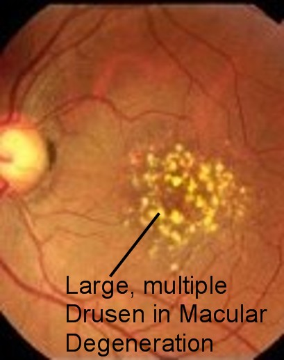
Drusen: Extracellular breakdown deposits (hyaline nodules/ colloid bodies)
2 types AMD
Dry (aka atrophic):.Drusen contained deep in Bruch's membrane
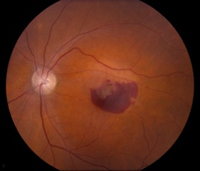
Wet (aka neovascularisation): Break in Bruch's membrane, abnormal vessels growing from choroid into retina
Aetiology
Unknown. ? inflammation in wet.
Pathogenesis
Deposits preventing exchange of nutrients and waste products between outer 1/3 of retina and choroid plexus
Risk Factors
Epidemiology
MD: Degenerative disease of the central portion of the retina (macula) that results primarily in loss of central vision.
Bruch's membrane: Thin membrane separating RPE (outermost layer of retina)/ retina from choroid
Drusen: Extracellular breakdown deposits (hyaline nodules/ colloid bodies)
2 types AMD
Dry (aka atrophic):.Drusen contained deep in Bruch's membrane
Wet (aka neovascularisation): Break in Bruch's membrane, abnormal vessels growing from choroid into retina
Aetiology
Unknown. ? inflammation in wet.
Pathogenesis
Deposits preventing exchange of nutrients and waste products between outer 1/3 of retina and choroid plexus
Risk Factors
- Age
- Family history
- Smoking
- Cardiovascular disease
Epidemiology
- Leading cause of blindness in elderly in developed countries
- 20% of dry develop into wet. 5% in 1 year, 15% in 3 years
- Wet: >40% chance can develop into both eyes

Clinical Features
Dry
Wet
Investigations
Imaging: OCT, FFA (fundus flourescine angiography), ICGA (indocyanine green angiography)
NOTE: ICGA's dye binds to albumin, provides better visualisation
NOTE: Angiography is to identify neovascularisation
Management
NOTE: All anti-VEGF treatments have similar efficacy. Avastin is used off-label for AMD
Source
www.uptodate.com 2013
www.opthobook.com 2009
CA Bradford, Basic Ophthalmology, 8th Ed 2004
Dry
- Usually asymptomatic
- Gradual vision loss
- RPE mottling (atrophy)
Wet
- Acute visual distortion (due subretinal haemorrhage/ oedema)
- Usually 1 eye but can be both
- Metamophorsia (distorting of Amsler Grid)
- Subretinal haemorrhage/ oedema/ exudate (not CMO cf PDR)
- Lack of fovea reflex
- Mottled appearance of RPE
Investigations
Imaging: OCT, FFA (fundus flourescine angiography), ICGA (indocyanine green angiography)
NOTE: ICGA's dye binds to albumin, provides better visualisation
NOTE: Angiography is to identify neovascularisation
Management
- Vitamin prophylaxis (Vit ACE, Zn, Cu --> Smokers cannot take A due to inc risk of Ca)
- Dry: Wait-and-see approach, Amsler Grid for progression
- Wet:
- Argon laser photocoagulation if not close to fovea
- Photodynamic therapy (PDT) with verteporfin if CNV beneath fovea
- Anti-VEGF treatment (Lucentis, Avastin, Aflibercept (Eylea))
NOTE: All anti-VEGF treatments have similar efficacy. Avastin is used off-label for AMD
Source
www.uptodate.com 2013
www.opthobook.com 2009
CA Bradford, Basic Ophthalmology, 8th Ed 2004
