
ECG
|
|
P wave = atrial depolarisation
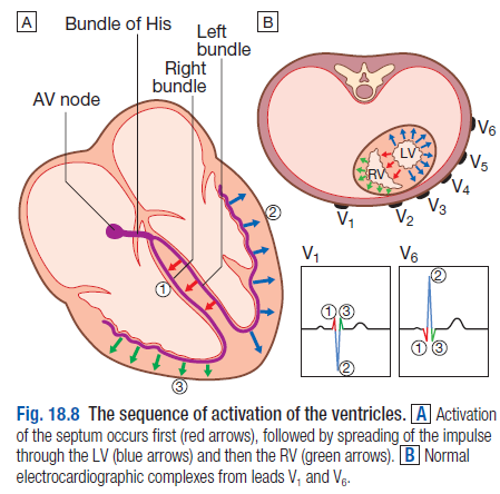
QRS = ventricular depolarisation
T wave = ventricular repolarisation
Normal
P wave < 3sm sq
PR 3-5 sm sq
QRS 1-3sm sq
QT < 10 sm sq (<0.42sm sq)
Anomalies
P waves
Delta waves
QRS
T waves
NOTE: normal female < 30y may see inverted T waves
QT interval
U waves
Other patterns
Source
Davidson's 22nd Ed 2014
www.geekymedics.com 2014
Dr Leovino Acharon 2014
QRS = ventricular depolarisation
T wave = ventricular repolarisation
Normal
P wave < 3sm sq
PR 3-5 sm sq
QRS 1-3sm sq
QT < 10 sm sq (<0.42sm sq)
Anomalies
P waves
- Tall = P pulmonale = R atrial enlargement
- Notched = P mitrale = L atrial enlargement
- No P waves + irregular = AF
- No P waves + normal QRS = SVT
- Short = WPW (p wave >3 sm sq + delta wave (slanted R wave) + shortened PR interval)
- Long = Heart block (1st deg = prolonged P waves; 2 mo 1 = PR inc then dropped narrow QRS, 2 mo 2 = PR constant + dropped wide QRS; 3rd deg independent p and QRS, both equally spaced from previous)
Delta waves
- WPW
QRS
- Short (<3 sm sq) = supraventricular origin
- Long duration (>3 sm sq) = ventricular origin = L/RBBB
- High amplitude = LVH
- Q wave (>1mm) = previous MI
- SVT = narrow complex tachy
- VT = left rabbit ear taller + Josephson's sign (notching on S nadir)
- VF = mess of ventricular activity
- Hypothermia < 32deg
- Depression (<1 sm sq from baseline) = Ischaemia, digoxin toxicity, tachy (anxiety) --> Base it on clinical context!!!
- Elevation = MI, pericarditis, LV aneurysm
- Reverse tick = digoxin effect (not toxicity)
T waves
- Tall (>5mm in precord, >10mm in std)= Infarct, Ischaemia, hyperK (tall tented) etc
- Inverted = hypoK,, tachy, ischaemia (similarly non-specific as ST depression)
NOTE: normal female < 30y may see inverted T waves
QT interval
- Long = hypoK, Mg, Ca, drugs, congenital long QT interval
U waves
- Hypokalaemia
Other patterns
- PE: T wave inversion V1-V3. S1Q3T3 (S - LI, Q - LIII, inverted T - LIII)
- LBBB: WiLliaM (W/V - V1; M - V6) + wide QRS (dominant S wave)
- RBBB: MaRroW (M - V1; W - V6) + wide QRS (dominant R wave)
- Posterior infarct: V1-V2 tall R waves; V1-V3 deep ST depression
- LVH: V1 + V5/6 > 7 large sq (<30y >8 large sq)
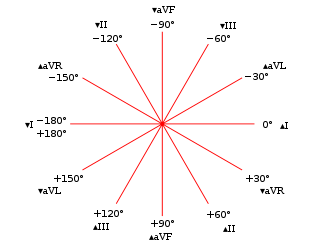
- LAFB: L axis deviation + qR complex lat leads + rS inferior leads (I and III deviation pattern)
- LPFB: R axis deviation + rS complex lat leads + qR complex inferior leads (I and III deviation pattern)
- Bifascicular block: RBBB + LAFB (more common)/ LPFB
- Trifascicular block: 1st deg AV block + RBBB + LAFB/LPFB
- Bigeminy: Every other beat is a premature atrial/junctional/ventricular Complex (PAC/ PJC/ PVC)
- Trigeminy: Every 3rd beat is a PAC/ PJC/ PVC
- Torsades de Pointes: Polymorphic VT "twisting" on an isoelectric line
Source
Davidson's 22nd Ed 2014
www.geekymedics.com 2014
Dr Leovino Acharon 2014
